Fig tree viruses in Morocco
Abstract
Fig (Ficus carica L.), widely grown in Morocco, is an important fruit crop for the country, both economically and socially. Fig mosaic disease is a complex with which at least eight viruses of different taxonomic position are associated. In 2014-2018, field surveys for viruses were carried out in the main fig growing areas of Morocco (Azilal, El Jadida, Moulay Driss Zarhoune and Taounate). A total of 117 samples were collected and checked by reverse transcription polymerase chain reaction (RT-PCR), using virus specific primers, for the presence of Fig mosaic virus (FMV), Fig leaf mottle-associated virus 1 (FLMaV-1), Fig leaf mottle-associated virus 2 (FLMaV-2), Fig mild mottling-associated virus (FMMaV), Fig latent virus 1 (FLV-1), Fig fleck-associated virus (FFkaV) and Fig cryptic virus (FCrV). PCR was also carried out for the presence of Fig badnavirus 1 (FBaV-1). About 71% of the trees were infected with at least one virus, with mixed infections in ca. 50% of the samples. FMV was the prevailing virus (40.9% infection), especially in Azilal (47.4%) and on cv. Nabout (46.2%), followed by FLMaV-1 (30.7%). FMMaV-2, FMMaV, FFkaV and FBV-1 were detected in 25.2%, 25.2%, 8.7% and 5.5% of the samples, respectively. FLV-1 and FCrV were not found. This is the first report on the presence of FMV, FLMaV-1, FLMaV-2, FMMaV, FFkaV and FBV-1 in Morocco and offers a preliminary insight into the unsatisfactory health status of fig trees in the country. Considering that the production of figs in Morocco is increasing, more attention should be given to improving the phytosanitary condition of fig trees in the country.
Keywords: Ficus carica, RT-PCR, virus detection, fig mosaic, Morocco
Introduction
The common fig (Ficus carica L.) is a temperate species native to South‐west Asia and Eastern Mediterranean region. It is widely cultivated throughout Morocco, mainly as individual trees in gardens and orchards for family consumption and only rarely as specialized crops, where fruits are used for fresh and dried consumption. The total Moroccan fig cultivated area is ca. 55,000 ha, with an estimated total production of 127,000 tonnes (Anonymous, 2017). Local cultivars, whose denominations often indicate the local geographic origin, the color of the fruits or their maturation period, are numerous and well adapted to agro-ecological Moroccan conditions, among which the most important are El Messari or Homrame or Johri, Lembdar Labied, Lembdar Lekhel, Rhoudane, El Koté and Aounq Hmam. The most important fig production areas are Taounate, Chefchaouen, Al Hoceima, Ouazzane and Tetouan.
Fig is affected to a very large extent by a disease known as “mosaic” (Fig mosaic disease, FMD), which is a serious pathological constraint of fig production. FMD remains one of the most serious pathological problem facing fig germplasm exchange and production. This disease, first reported from California (Condit and Horne, 1933), is now known to have a worldwide distribution, likely being present in all countries were fig is grown (Blodgett and Gömec, 1967; Martelli et al. 1993). FMD is a graft transmissible disease (Condit and Horne, 1933) vectored by the eriophyid mite Aceria ficus (Flock and Wallace, 1955; Slykhuis, 1973). Although no estimates of the economic impact of FMD are available, the notion that severely affected trees are less productive than those with milder symptoms and suffer premature fruit abscission has been taken as an indication that FMD can have a detrimental effect on the crop (Chiumenti et al., 2013). FMD is a complex disorder (Martelli, 2011) with which eight viruses of different taxonomic position are associated (Table 1). Fig mosaic virus (FMV) is the agent that occurs in symptomatic plants more often than any of the other fig-infecting RNA viruses and is the major incitant of mosaic (Chiumenti et al., 2013). The role in symptom induction of other viruses, such as the putative closterovirids Fig leaf mottle-associated virus 1 (FLMaV-1), Fig leaf mottle-associated virus 2 (FLMaV-2), Fig mild mottle-associated virus (FMMaV) (Elbeaino et al., 2006, 2007, 2010) and the putative marafivirus Fig fleck-associated virus (FFkaV) (Elbeaino et al., 2011a), has not been ascertained. None of the aforementioned viruses is transmitted through seeds, contrarily to Fig latent virus 1 (FLV-1) (Gattoni et al., 2009) and Fig badnavirus 1 (FBV-1), the only DNA virus identified in fig so far (Laney et al., 2012; Minafra et al., 2012), both of which are vertically transmitted to seedlings, in which they do not induce symptoms. Two other closteroviruses, Arkansas fig closterovirus 1 and 2 (AFCV-1 and -2), and the badnavirus FBV-1 were also reported in Arkansas (Tzanetakis and Martin, 2010) with evidence that FBV-1 is a DNA virus that is integrated in the fig genome (Laney et al., 2012).
Foliar discolorations (chlorotic mottling, blotching and banding, clearing and feathering of the veins, chlorotic and necrotic ringspots and line patterns) and malformation resembling those typical of fig mosaic disease (Martelli, 2011) had been repeatedly observed in Moroccan fig orchards. However, no information is currently available on the possible causal agents. Accordingly, the main objective of the present study was to investigate and evaluate the incidence and distribution in Moroccan fig orchards of the following viruses: FMV, FLMaV-1, FLMaV-2, FMMaV, FLV-1, FFkaV and FCrV and FBV-1.
Material and methods
During 2014-2018, field surveys were carried out in 32 commercial fig plantations located in four Moroccan regions: Azilal, El Jadida, Moulay Driss Zarhoune and Taounate. Samples were representative of the most common local fig varieties grown in Morocco, i.e. Aounq, Boustati, Elquoti Lebied, El Messari, Embar Lekhel, Chetoui, Jeblia, Lembdar Lebied, Lembdar Lekhel, M’tioui, Nabout and Rhoudane. In total, 127 samples were randomly collected from symptomatic (71 samples) and symptomless (56 samples) fig trees. Selection of orchards and collection of samples was done according to the prevalence of the cultivars and their geographical distribution. Samples consisting of leaves and cuttings of about 30 cm in length were collected from one- to two-year-old shoots from the quadrant of the tree canopy and stored in plastic bags at 4°C until use for laboratory assays.
Total nucleic acids (TNAs) were extracted from 100 mg of leaf veins or cortical scrapings of fig samples using “silica capture” method as described by Foissac et al. (2001). Ten μl of TNAs were denatured by boiling at 95°C for 5 min, then reverse‐transcribed (with the exception of FBaV‐1) with random primers and M‐MLV reverse transcriptase (Invitrogen Laboratories, USA) for 1 h at 39°C (Minafra and Hadidi, 1994). The amplification was performed using 2.5 μl cDNA (TNAs for FBaV‐1), in a total volume of 25 μl containing 2.5 μl of 10X Taq polymerase buffer, 0.5 μl of 50 mM MgCl2, 0.5 μl of 10 mM dNTPs, 0.5 μl of 10 μM primer (sense), 0.5 μl of 10 μM primer (anti-sense) and 0.25 μl of GoTaq polymerase (5 unit/μl) (Promega Company, USA).
FMV, FLMaV-1, FLMaV-2, FMMaV, FLV-1, FFkaV, FCrV and FBV-1 were detected (Table 1) using sets of specific primers (Table 2). PCR amplifications were carried out using an initial denaturation at 94°C for 4 min, followed by 35 cycles at 94°C for 30 sec, annealing at 58°C (55°C for FLV-1) for 35 sec, and extension 72°C for 30 sec. Final elongation was carried out at 72°C for 7 min. Ten μL of the PCR reactions were electrophoresed in 1.2% agarose gel in 1X TAE buffer and stained with ethidium bromide.
| Virus species | Genus | Reference |
| Fig mosaic virus (FMV) | Emaravirus | Elbeaino et al., 2009 |
| Fig leaf mottle associated virus 1 (FLMaV-1) | Closterovirus | Elbeaino et al., 2006 |
| Fig leaf mottle associated virus 2 (FLMaV-2) | Ampelovirus | Elbeaino et al., 2007 |
| Fig mild mottle-associated virus (FMMaV) | Closterovirus | Elbeaino et al., 2010 |
| Fig fleck-associated virus (FFkaV) | Maculavirus | Elbeaino et al., 2011a |
| Fig latent virus 1 (FLV-1) | Trichovirus | Gattoni et al., 2009 |
| Fig cryptic virus (FCrV) | Alphacryptovirus | Elbeaino et al., 2011b |
| Fig badnavirus 1 (FBV-1) | Badnavirus | Laney et al., 2012 |
Table 2. PCR primers used in the present study.
| Virus species | Primer | Sequence (5’-3’) | Amplicon size (bp) | References |
| FMV | BB42 up | TGGCAGATTCAAGGATAATGG | 218 | Elbeaino et al., 2009 |
| BB42 down | TGGGACATTCTTGTGTCAGG | |||
| FLMaV-1 | N17s | CGTGGCTGATGCAAAGTTTA | 350 | Elbeaino et al., 2006 |
| N17a | GTTAACGCATGCTTCCATGA | |||
| FLMaV-2 | F3s | GAACAGTGCCTATCAGTTTGATTTG | 360 | Elbeaino et al., 2007 |
| F3a | CCCACCTCCTGCGAAGCTAGAGAA | |||
| FMMaV | LM3s | AAGGGGAATCTACAAGGGTCG | 311 | Elbeaino et al., 2010 |
| LM3a | TATTACGCGCTTGAGGATTGC | |||
| FLV-1 | FFup | CGCTTTGCCCCAATGTGCAGAT | 125 | Gattoni et al., 2009; Modified by Chiumenti et al., 2013 |
| FFrev25 | TARTCDGATTCHACRCACAGGTC | |||
| FBV-1 | P1-s | GCTGATCACAAGAGGCATGA | 214 | Minafra et al., 2012 |
| P1-as | TCCTTGTTTCCACGTTCCTT | |||
| FFkaV | d8-s | ATGACGACTGTCAACTCCCT | 270 | Elbeaino et al., 2012a |
| d8-a | TTAAGCCAGGGTGGGAGTGTTG | |||
| FCrV | R1-s | TCGATTGTCTTTGGAGAGG | 353 | Elbeaino et al., 2011b |
| R1-a | CGCATCCACAGTATCCCATT |
Results and discussion
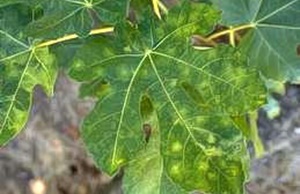
Fig mosaic symptoms were observed in most of the surveyed fields, on young and old leaves, especially during spring and autumn. Trees showed a wide array of leaf discolorations, i.e. chlorotic to yellowish mottling and blotching, mosaic spots, vein clearing and feathering, chlorotic and necrotic ringspots. Leaf malformations were also encountered during the surveys and were associated with discolorations and chlorotic mottling with a contrast ranged from yellow to green color.
RT-PCR results (PCR for FBV-1) showed that 91 samples, out of 127 tested, were infected by at least one virus, which represents an infection rate of 71.6% (Table 3). Single infections accounted for 25.2% (32/127) of the samples collected from plants. Mixed virus infections were common involving two (23.6%), three (18.9%) or four viruses (2.4%) in various combinations. No plant was found to be infected by more than four viruses encountered in the survey. Results showed the highest incidence for FMV (40.9%), followed by FLMaV-1 (30.7%), FLMaV-2 (25.2%) and FMMaV (25.2%) among the collected samples. FFkaV and FBV-1 were detected in 8.7 and 5.5% of the samples, respectively. FCrV and FLV-1 were not detected. The prevalence of the above viruses in fig trees varied according to the region. The highest infection rate was observed in Taounate (82.7%), followed by Moulay Driss Zarhoune (68.2%), Azilal (63.1%) and El Jadida (61.8%). The infection rate was high in commercial cvs. Lembdar Lekhel (100%), Lembbdar Lebied (88.9%), El Messari (81.8%), Rhoudane (75%) and El quoti Lebied (72.7%).
Results showed that six viruses, characterized in this study, were present in the main Moroccan fig-growing areas, with levels of infections that were substantially in line with those reported from other Mediterranean countries with few exceptions. In particular, the presence of FMV in Moroccan fig trees was high (40.9%) when compared to previous reports in Turkey (8.6%) (Caglar et al., 2011) and Tunisia (34.5%) (El Air et al., 2015), but lower than that reported in Lebanon (42.2%) (Elbeaino et al., 2012b) and Syria (56.7%) (Elbeaino et al., 2012a). The incidence of FLMaV-1 was lower (30.7%) than that reported in Lebanon (36.3%) (Elbeaino et al., 2012b), but higher than that reported in Tunisia (14.8%) (El Air et al., 2015). The incidence of FLMaV-2 was lower (25.2%) than that reported in Lebanon (29.4%) and Syria (31.1%) (Elbeaino et al., 20012a,b), but much higher that reported in Tunisia (4.3%) (El Air et al., 2015). FMMaV was widely distributed in Morocco (25.2%) when compared to previous reports from Turkey (2%) (Caglar et al., 2011), Syria (12.2%) (Elbeaino et al., 2012a) and Tunisia (10.7%) (El Air et al., 2015). The Incidence of FFkaV (8.7%) was lower than that recorded from other Mediterranean countries such as Tunisia (10.3%), Lebanon (13.7%) and Syria (36.7%) (El Air et al., 2015; Elbeaino et al., 2012a,b). Although FBV-1, the only DNA virus found in fig, is vectored primarily by mealybugs and aphids (Jones et al., 2002), it was showed to be the less widespread virus in all cultivars. FBV‐1 was found to infect all different F. carica organs (syconium, leaf and bud) which confirms its vertical transmission to seedlings and its hypothesized integration in the host genome (Laney et al., 2012).
Of the 56 trees that did not show apparent FMD-like symptoms at the time of the survey, 31 were PCR-negative for FMV but contained other viruses they were tested for, supporting the complex nature of FMD, in whose aetiology FMV plays a significant but likely not an exclusive role (Martelli, 2011). Of the 71 symptomatic plants, 57 (80.3%) were FMV-positive, thus confirming the high level of the association between FMD and FMV (Martelli, 2011). During a preliminary survey in the Canary Islands in autumn of 2009, Elbeaino et al. (2011c) found that among 12 trees that did not show apparent FMD-like symptoms at the time of the survey, eight were FMV-negative, whereas 15 (88%) of the 17 symptomatic plants were FMV-positive.
Interestingly, at least one tree of each of the surveyed 12 cultivars did not show visible symptoms at the time of the survey and did not contain any of the eight viruses. These plants, after a confirmatory round of additional assays, may represent potential sources of material for propagation in the framework of a sanitary improvement programme, which has been already established since 2013 in Morocco.
Table 3. Viruses detected in fig trees in Morocco.
| Region | Sampled groves (No.) | Cultivar | Tested trees No. | Infected trees | FMV | FLMaV-1 | FLMaV-2 | FMMaV | FFkaV | FBV-1 | |||||||
| No. | % | No. | % | No. | % | No. | % | No. | % | No. | % | No. | % | ||||
| Azilal | 6 | Chetoui | 9 | 6 | 66.7 | 4 | 44.4 | 3 | 33.3 | 2 | 22.2 | 3 | 33.3 | - | 0 | 1 | 11.1 |
| Elquoti Lebied | 6 | 3 | 50 | 3 | 50 | 2 | 33.3 | 1 | 16.7 | - | 0 | - | 0 | - | 0 | ||
| Rhoudane | 4 | 3 | 75 | 2 | 50 | 2 | 50 | 2 | 50.0 | 1 | 25 | - | 0 | - | 0 | ||
| El Jadida | 8 | Nabout | 13 | 8 | 61.5 | 6 | 46.2 | 4 | 30.8 | 3 | 23.1 | 3 | 23.1 | 1 | 7.7 | 1 | 7.7 |
| Embar Lekhel | 9 | 5 | 55.5 | 4 | 44.4 | 2 | 22.2 | - | 0 | 2 | 22.2 | 1 | 11.1 | - | 0 | ||
| M’tioui | 12 | 8 | 66.7 | 6 | 50 | 2 | 16.7 | 1 | 8.3 | 1 | 8.3 | 1 | 8.3 | 1 | 8.3 | ||
| Moulay Driss Zarhoune | 5 | Bousbati | 10 | 7 | 70 | 4 | 40 | 2 | 20 | 3 | 30 | 1 | 10 | - | 0 | - | 0 |
| Embar Lekhel | 12 | 8 | 66.7 | 2 | 16.7 | 3 | 25 | 4 | 33.3 | 3 | 25 | 1 | 8.3 | - | 0 | ||
| Taounate | 13 | Jeblia | 11 | 7 | 63.6 | 4 | 36.4 | 4 | 36.4 | 3 | 27.3 | 3 | 27.3 | 1 | 9.1 | 1 | 9.1 |
| El Messari | 11 | 9 | 81.8 | 4 | 36.4 | 2 | 18.2 | 3 | 27.3 | 3 | 27.3 | 3 | 27.3 | 2 | 18.2 | ||
| Lembdar Lebied | 9 | 8 | 88.9 | 4 | 44.4 | 4 | 44.4 | 3 | 33.3 | 2 | 22.2 | 1 | 11.1 | - | 0 | ||
| Lembdar Lekhel | 9 | 9 | 100 | 4 | 44.4 | 3 | 33.3 | 3 | 33.3 | 5 | 55.6 | - | 0 | - | 0 | ||
| Aounq | 7 | 5 | 71.4 | 3 | 42.9 | 2 | 28.6 | 1 | 14.3 | 3 | 42.9 | 1 | 14.3 | - | 0 | ||
| Elquoti Lebied | 5 | 5 | 100 | 2 | 40 | 4 | 80 | 3 | 60 | 2 | 40 | 1 | 20 | 1 | 20 | ||
| Total | 32 | 127 | 91 | 71.6 | 52 | 40.9 | 39 | 30.7 | 32 | 25.2 | 32 | 25.2 | 11 | 8.7 | 7 | 5.5 |
Conclusion
Fig mosaic disease induces the major threat to the fig crop and may constitute a limiting factor for its growing. The present study expands the knowledge on the sanitary status of fig trees in Morocco and provides further information about the virus incidence and distribution within the country. The results have shown a much deteriorated sanitary status of the fig crop in Morocco (71.6% of viral infections). All the tested fig‐infecting viruses were present in the surveyed cultivars with the exception of FCrV and FLV‐1. These results are not surprising considering the mode of propagation of this species (by rooted cuttings and grafting) and the presence of very efficient virus vectors (eriophyid mites, mealybugs and aphids), both factors that favour the transmission of viral agents in nature. The knowledge gained in recent years on virus diseases of fig can finally allow initiating the sanitary selection and sanitation of propagating material.
References
Anonymous. (2017). La filière du figuier: Entre pari de valorisation et défi de commercialisation. http://www.agrimaroc.ma/figuier-valorisation-commercialisation/
Blodgett E.C. and Gömec C. (1967). Fig mosaic. Plant Disease Reports 51: 893-896.
Caglar B.K., Fidan H., Guldur M.E. and Elbeaino T. (2011). The prevalence of three viruses infecting fig in southern Turkey. Journal of Phytopathology 159: 181–183.
Chiumenti M., Campanale A., Bottalico G., Minafra A., De Stradis A., Savino V. and Martelli G.P. (2013). Sanitation trials for the production of virus-free fig stocks. Journal of Plant Pathology 95: 655-658.
Condit I.J. and Horne W.T. (1933). A mosaic of fig in California. Phytopatology 23: 887-897.
El Air M., Mahfoudi N., Digiaro M., Dhouibi M.H. and Elbeaino T. (2015). Incidence and distribution of viruses in Tunisian fig orchards. Journal of Plant Pathology 97: 327-331.
Elbeaino T., Abou Kubaa R., Ismaeil F., Mando J. and Digiaro M. (2012a). Viruses and Hop stunt viroid of fig trees in Syria. Journal of Plant Pathology 94: 687-691.
Elbeaino T., Mortada C., Digiaro M. and Choueiri M. (2012b). Survey of fig viruses in Lebanon. Proc. XXVIIIth IHC-IS on the Challenge for a Sustainable Production, Protection and Consumption of Mediterranean Fruits and Nuts. In: D’Onghia A.M. et al., (eds.). Acta Hort. 940: 665-668.
Elbeaino T., Digiaro M. and Martelli G.P. (2011a). Complete nucleotide sequence of Fig fleck-associated virus, a novel member of the family Tymoviridae. Virus Research 16: 198-202.
Elbeaino T., Abou Kubaa R., Digiaro M., Minafra A. and Martelli G.P. (2011b). The complete nucleotide sequence and genome organization of Fig cryptic virus, a novel bipartite dsRNA virus infecting fig, widely distributed in the Mediterranean basin. Virus Genes 42: 415-421.
Elbeaino T., González-Rodríguez Á.M., Grajal-Martín M.J. and Digiaro M. (2011c). Survey of fig viruses in the Canary Islands. Journal of Plant Pathology 93: 737-739.
Elbeaino T., Digiaro M., Heinoun K., De Stradis A. and Martelli G.P. (2010). Fig mild mottle-associated virus, a novel closterovirus infecting fig. Journal of Plant Pathology 92: 165-172.
Elbeaino T., Digiaro M., Alabdullah A., De Stradis A., Minafra A., Mielke N., Castellano M. A. and Martelli G.P. (2009). A multipartite single-stranded negative-sense RNA virus is the putative agent of fig mosaic disease. Journal of General Virology 90: 1-8.
Elbeaino T., Digiaro M., De Stradis A. and Martelli G.P. (2007). Identification of a second member of the family Closteroviridae in mosaic-diseased figs. Journal of Plant Pathology 89: 199-124.
Elbeaino T., Digiaro M., De Stradis A. and Martelli G.P. (2006). Partial characterization of a closterovirus associated whit a chlorotic mottling of fig. Journal of Plant Pathology 88: 187-192.
Foissac X., Svanella-Dumas L., Dulucq M. J., Candresse T. and Gentit P. (2001). Polyvalent detection of fruit tree tricho, capillo and foveavirus by nested RT-PCR using degenerated and inosine containing primers (DOP RT-PCR). Acta Horticulturae 550: 37-44.
Flock R.A. and Wallace J.M. (1955). Transmission of fig mosaic by the eriophyd mite Aceria ficus. Phytopathology 45: 52-54.
Gattoni G., Minafra A., Castellano M.A., De Stradis A., Boscia D., Elbeaino T., Digiaro M. and Martelli G.P. (2009). Some properties of Fig latent virus 1, a new member of the family Flexiviridae. Journal of Plant Pathology 91: 555-564.
Jones A.T., McGavin W.J., Geering A.D.W. and Lockhart B.E.L. (2002). Identification of Rubus yellow net virus as a distinct badnavirus and its detection by PCR in Rubus species and in aphids. Ann. Appl. Biol. 141: 1-10.
Laney A.G., Hassan M. and Tzanetakis I.E. (2012). An integrated badnavirus is prevalent in fig germplasm. Phytopathology 102: 1182-1189.
Martelli G.P., Castellano M.A. and Lafortezza R. (1993). An ultrastructural study of fig mosaic. Phytopathologia Mediterranea 32: 33-43.
Martelli G.P. (2011). Fig mosaic disease and associated pathogens. In: Hadidi A., Barba M. Candresse T. and Jelkmann W. (eds). Virus and Virus-like Diseases of Pome and Stone Fruits. APS Press, St. Paul, MN, USA, pp. 281-287.
Minafra A., Chiumenti M., Elbeaino T., Digiaro M., Bottalico G., Pantaleo V. and Martelli G.P. (2012). Occurrence of Fig badnavirus 1 in fig trees from different countries and in symptomless seedlings. Journal of Plant Pathology 94: S4.105.
Minafra A. and Hadidi A. (1994). Sensitive detection of Grapevine virus A, B and leafroll-associated virus III from viruliferous mealybugs and infected tissues by cDNA amplification. Journal of Virological Methods 47: 175-187.
Slykhuis J.T. (1973). Viruses and mites. In: Gibbs A.J. (eds). Viruses and Invertrebates, pp. 391-405. North Holland Publishing Co., Amsterdam, The Netherlands.
Tzanetakis I. and Martìn R. (2010). New viruses found in fig exhibiting mosaic symptoms. 21st International Conference on Virus and other Graft Transmissible Diseases of Fruit Crops. Julius-Kühn-Archiv: 427.