Evaluation of the toxicity of aqueous extract of the Ganoderma applanatum mushroom
Abstract
Plants and fungi, are widely used around the world as a source of traditional medicine and human food. Nowadays, scientists and nutritionists encourage the use of plants and fungi although cases of poisoning are frequently reported. Many countries around the world, including the Democratic Republic of the Congo, use Ganoderma applanatum mushroom for its proven therapeutic virtues. However, the people of Sankuru have often suspected it of being poisonous. The purpose of this study was therefore to evaluate the supposed toxicity of this mushroom. For this, a chemical screening was carried out in test tubes containing methanolic and aqueous extracts, followed by a screening by thin layer chromatography in order to detect the presence of secondary metabolites. The aqueous extract was administered to mice at rates ranging from 5000 to 1000 mg/kg to assess the toxicity of the G. applanatum sample. Examination of Alanine amino-transferase (ALAT) and Asparate amino-transferase (ASAT) in the mice blood showed no resistance and no alteration of vital organs (liver and heart) even at the highest rate (5000 mg/kg). The results of the chemical screening revealed the presence of various compounds, including alkaloids, terpenes, flavonoids, phenols and others. The administration of the different doses did not cause any lethality to the animals. The results of this study argue in favor of the safety of Ganoderma applanatum in the traditional treatment of certain diseases by oral route.
Keywords: Ganoderma applanatum, traditional use, chemical compounds, Sankuru, Democratic Republic of the Congo
INTRODUCTION
Mushrooms are widely used around the world as a source of traditional medicine and human food (Diansambu et al., 2015). The food interest of mushrooms is often sought because of their aromas appreciated by gourmets (Scholer and Boer 2018; Tejedor et al., 2023). Many data published around the world emphasize the high nutrient value of edible mushrooms. They are considered as a source of lipids, carbohydrates, proteins, vitamins and minerals (Wani et al., 2010; Wandati et al., 2013). Moreover, apart from fruits and vegetables, mushrooms are known to contain active compounds in antioxidant and anticancer properties (Kozarski et al., 2012; Elkhateeb et al., 2018; Kumar et al., 2021).
The Democratic Republic of the Congo is among the countries of Sub-Saharan Africa that consume mushrooms. This foodstuff is very popular with the Congolese population, not only to alleviate the recurring problem of food insecurity, but also for their use in traditional medicine in the treatment of many pathologies (Ishara et al., 2018; Kamalebo et al., 2018).
However, the consumption of mushrooms is not always without risk and some of them can cause poisoning due to either their own toxins (mycetism), the development of micro-organisms, or a polluted harvesting site. The spores produced in large numbers can also cause allergies to some people (Barceloux, 2008; Rapior and Fons, 2011). Some species of mushrooms may also show toxicity if undercooked (Boels et al., 2014).
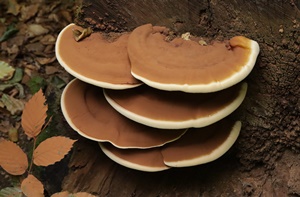
Ganoderma applanatum is a species of fungi belonging to the genus Ganoderma, widely distributed in the world (Luo et al., 2016; Jeong et al., 2018). It is widely used in traditional Asian medicine because of its pharmacological properties and as food (Guillamon et al., 2010; Wasser, 2011; Akpera et al., 2011; Shahrajabian et al., 2019). Although considered an important source of medicine and frequently used as an extract by decoction in traditional medicine, information gathered from the local population of the province of Sankuru in the Democratic Republic of the Congo shows that this species of mushroom is toxic. With this in mind, this study was initiated to highlight the pharmacological properties of this species.
MATERIAL AND METHODS
Biological material
The samples of Ganoderma applanatum were collected from Lodja, the chief city of the province of Sankuru, in the Democratic Republic of Congo. The taxonomic identification of the G. applanatum was done by the Mycology laboratory of the Biology, Department of the Faculty of Sciences, University of Kinshasa.
Preparation of G. applanatum mushroom powder
The samples of collected G. applanatum were dried in the shade and crushed using a mortar and pestle. Then the mushroom powder was obtained using a sieve.
Extraction and chemical screening
Chemical screening was performed based on the method recently described by Mpiana et al. (2012) and Ngbolua et al. (2015). The mushroom powder (10 mg) was macerated in 50 ml of distilled water (aqueous extract) and in methanol (organic extract) for 24 hours, so as to carry out the phytochemical screening in solution. Each extract obtained after maceration using an ultrasonic bath was used for thin layer chromatography (TLC). This aqueous extract obtained after infusion of the evaporated powder at 45°C was used to evaluate the bioactivities. To do this, we weighed 2 g of the powder using a sensitive scale, then added 50 ml of distilled water to reconstitute the total aqueous extract of G. applanatum mushrooms. For comparative analysis, different mushroom extracts were prepared. Then, 5 g of mushroom powder were successively extracted with 125 ml of ethanol, 125 ml of ethyl acetate and 125 ml of hexane.
Chemical screening of the aqueous extract
Detecting of the total polyphenols
They are subdivided into several families including flavonoids and anthocyanins (Muanda, 2010; Bruneton, 1999).
The analysis of polyphenols is carried out using Burton’s reagent, which is a mixture of FeCL3 2% and K3Fe(CN)6 1% (1:1, V/V). We introduced 3 ml of aqueous extract into a test tube and added 1ml of Burton’s reagent. In the presence of polyphenols, the solution turns intense blue and is sometimes accompanied by a precipitate. In case of a positive test, we then systematically looked for the various polyphenolic compounds such as flavonoids, quinones, anthocyanins, tannins, leuco anthocyanins, etc...
Flavonoids are compounds with large carbon chains (15 atoms) to which are grafted benzene rings united to a small chain of 3 carbon atoms (Panche et al., 2016). This assay is done using Shinoda’s reagent, consisting of a mixture of 95% ethanol, concentrated HCl, distilled water (1:1:1 v/v/v) and Mg or Zn shavings.
We placed 3 ml of organic extract in the test tube and then added a few drops of Shinoda’s reagent using a Pasteur pipette a few shavings of Mg (effervescence is observed) and a few drops of isoamyl alcohol. We stirred the mixture and let it until settles. The formation of a thin film in the supernatant layer of isoamyl alcohol indicates the presence of:
• Flavones: if the color is red to reddish-orange;
• Flavonols: if the color is cherry red;
• Flavonones: if the color is purplish red.
Detecting of bond quinones
Quinones are compounds corresponding to the oxidation of aromatic compounds (phenols for example) and characterized by a 1,4-diketo-2,5-cyclohexadiene (p-quinone) unit or possibly by a 1,2-diketo-cyclohexadiene-3.5 (o-quinone) (Panche et al., 2016). This analysis is done using Borntrager’s reagent (10% NaOH or 10% NH4OH).
We took 3 ml of the aqueous extract and placed it in a test tube and added Borntrager’s reagent and stirred vigorously. The appearance of a color ranging from orange to bright red is a positive test.
Anthocyanins are water-soluble pigments that color certain plant flowers blue, red, purple, pink or orange. They belong to the family of total polyphenols (Hopkins, 2003). The detection of anthocyanins is done using concentrated hydrochloric acid (HCl) concentrated at 20%.
We introduced 3 ml of aqueous extracts in a test tube, then added a few drops of 20% HCl reagents, and heated it in a water bath for 20 minutes. In the presence of anthocyanins, a purplish color develops there due to the formation of anthocyanin chloride which can crystallize.
Detecting leuco-anthocyanin
The detection of these compounds (leuco-anthocyanins) is done using Shinoda’s reagent.
We placed 3 ml of aqueous extract in a test tube, then added a few drops of Shinoda’s reagent and a few drops of iso-amyl alcohol, and heated the mixture in a water bath. The presence of a red or purplish color in the supernatant layer is a positive test.
Detecting of tannins
Tannins are water-soluble phenolic compounds which exhibit, alongside the classic reactions of phenols, the properties of precipitating proteins and alkaloids. These are known as hydrolysable tannins (gallic tannins and catechin tannins). They are found in the leaves, barks and even in the flowers of certain plants and fungi (Praveen and Kumud, 2012). They are detected using Stiansny’s reagent (formaldehyde 30% + concentrated HCl 2:1/v:v) and FeCl3 20%, Burton’s reagent.
We placed 2.5 ml of aqueous extract and then added 1.0 ml of FeCl3 2% and 1.0 ml of Stiansny’s reagent in a test tube. The presence of a red-greenish color, with or without a precipitate, indicates the presence of tannins in general.
In case of a positive test, one can differentiate gallic tannins from catechin tannins according to the following procedure:
• Take 2.5 ml of aqueous extract in a test tube, add 2 ml of Stiansny’s reagent and heat it in a bain-marie at 90°C for 30 minutes. A brown precipitate indicates the presence of catechin tannins.
• If catechin tannins are present, filter the mixture and saturate the filtrate with crystals of sodium acetate (CH3COONa).
• After adding 1.0 ml of FeCl3 2%, a blackish color indicates the presence of water-soluble tannins or gallic tannins.
Detecting of alkaloids
Alkaloids are nitrogenous compounds, generally heterocyclic, with more or less pronounced alkaline reactions and endowed with marked pharmacodynamic properties at low doses (Bouchelta et al., 2009). They are detected using Dragendorff’s reagent and/or Mayer’s reagent (potassium mercurotetraiodide).
With Dragendorff’s Reagents
We placed 3 ml of aqueous extract in a test tube, slightly acidified with 1 ml of HCl 0.1 N and a few drops of Dragendorff’s reagent. The appearance of a red-orange precipitate indicates a positive test (presence of alkaloids in the solution).
With Mayer Reagents
We weighed 3.0 g of the powder/sample and macerated them in 3 ml of 5% HCl solution. We stirred for 30 minutes with the vortex, left the solution to stand and then filtered. After filtration, we put 1 ml in a test tube and added three drops of Mayer’s reagent. The presence of cloudiness in the solution or the formation of a precipitate in the solution confirms the presence of alkaloids.
Search of Saponins
Saponins or saponosides are glycosides with steroidal or tri-terpenics characterized by their surfactant properties. They form a persistent foaming solution when dissolved in water (Hopkins, 2003).
We put 3 ml of aqueous extract in the test tube, which we added 1 ml of distilled water and then we shaked vigorously. The formation of a resistant foam at least 1 cm high for 15 minutes indicates the presence of saponins or a positive test.
Chemical screening on the organic extract
Research of steroids and terpenes
Steroids can be considered as tetracyclic triterpenes having lost at most the cyclopentanonperhydrophenanthrenic structure, while terpenes (triterpenoids) are natural derivatives of squalene with an always polycyclic structure, currently tetracyclic and slightly acyclic (Salvador et al., 2009).
The detection of these two families of compounds is carried out using the Leibermann-Burchardat reagent which composition is the mixture of acetic anhydride (CH3CO)2O and concentrated sulfuric acid (H2SO4) 97%.
We put 5ml of extract in a test tube and evaporated it to dryness. Add Leibermann-Burchardat reagent. A purple color indicates the presence of triterpenoids and steroids in a mixture. Separately, the triterpenoids give a mauve complex, while steroids, on their side, develop a green color.
Thus during the titration, the mauve colored solution indicates the presence of triterpenoids (and/or terpenes). However, the green staining solution is a positive test for steroids.
Detecting of tannins
These compounds are detected using Stiansny’s reagent and Burton’s reagent.
Extract with ethyl acetate
We took 3 ml of the organic extract in a test tube. Then we added three drops of Stiansny’s reagent. A color change or formation of a greenish-black precipitate indicates that the test is positive for gallic tannins.
Extract from the general flavonoid test (Cfr. CCM)
We took 2 ml of the extract obtained in the general test of flavonoids, then added a few drops of Burton’s reagent (solution of Fe Cl3 1% and K3 Fe (CN)6 1%). A dark blue, dark green, or black color is a positive test for tannins.
We took 3 ml of extract the solution obtained in the general flavonoid test, then added 1.5 ml of Stiansny’s reagent (formaldehyde 30% HCl concentrated 2:1). Heating in a water bath at 90°C causes the quantitative precipitation of catechin tannins (pink precipitate).
After filtration, the filtrate saturated with sodium acetate is added with a few drops of Burton’s reagent. A blue or black tint indicates the presence of gallic tannins.
Detecting of free quinones
It is done using Borntrager’s reagent (NaOH 10% or NH4OH 10%).
We took 3 ml of extract in a tube, then added a few drops of Borntrager’s reagent and shake vigorously. The appearance of a color ranging from orange to bright red is a positive test.
Screening by thin layer chromatography (TLC)
Thin layer chromatography (TLC) was performed following the standard protocol described by Poole (2003) based on the observation of spots of various colors to identify different secondary metabolites.
Sample preparation
1 g of G. applanatum powder mixed with 10 ml of dichloromethane for 15 minutes with thermal stirring. The filtrate was evaporated to dryness and the residue was dissolved in 0.5 ml of toluene which served for the identification of flavonoids, coumarins, triterpenes and anthracene derivatives.
Flavonoids
Chromatographic conditions
Stationary phase: Silica gel F254;
Mobile phase: ethyl acetate-formic acid-glacial acetic acid-water (50:6.5:6.5:13.5);
Revelation: once developed, the chromatoplate is observed under UV at 366 nm, then the Neu reagent is sprayed and followed by observation under UV at 366 nm. The presence of flavonoids is visualized by blue fluorescent spots.
Coumarins
Chromatographic conditions
Stationary phase: Silica gel F254;
Mobile phase: toluene-ethyl acetate (31:2.3, saturated with 10% acetic acid);
Revelation: was carried out under UV at 366 nm. The blue color was characteristic of coumarins.
Triterpenes
Chromatographic conditions
Stationary phase: Silica gel F254;
Elution system (mobile phase): toluene-ethyl acetate (31:2,3);
Revelation: sulfuric anisaldehyde and heating for 10 minutes at 100°C;
Detection: Terpenes give spots of blue color after revelation in the visible.
Anthracene drifts
Chromatographic conditions
Stationary phase: Silica gel F254;
Mobile phase: ethyl acetate-methanol-water (50: 8.5: 6.5);
The revelation was carried out under UV at 366 nm and the plate was sprayed with 10% KOH in ethanol;
Detection: the anthracene derivatives give fluorescent spots of red color at 366 nm.
Alkaloids
Sample preparation
1g of mushroom powder was macerated in 1 ml of 10% ammonia or 10% Na2CO3, 5 ml of ethyl acetate were added and the whole is left under thermal stirring for 30 minutes. 10 μl of the filtrate was used for TLC analysis.
Chromatographic conditions
Stationary phase: Silicagel F254;
Mobile phase: toluene - ethyl acetate - ethylamine (35:10:5);
Development: The 5% NaNO2 reagent was used as a visible light developer;
Detection: The yellow spots denote the presence of alkaloids.
Toxicity test of Ganoderma applanatum mushroom extracts
Preparation of the total aqueous extract of Ganoderma applanatum
The total aqueous extract was prepared on the basis of G. applanatum mushroom powder, a method recommended in traditional medicine (decoction) (Konkon et al., 2006). For this experimental study, the methods were adapted to the reality of the laboratory. For this, 100 g of mushroom powder were added to 2 liters of distilled water in a suitable container (metal pot). Everything was boiled for 15 minutes. The obtained solution was filtered and the filtrate was evaporated in an oven at a temperature of 50°C. The brown colored dry extract obtained constitutes the total aqueous extract of the G. applanatum mushrooms.
Acute oral toxicity
This experimental study was adapted to that described by guideline 423 (OECD, 2001). The concentrations of the total aqueous extract of G. applanatum were prepared based on the principle that the concentrations to be administered should be related to the body weight of the mice. Thus, the doses were expressed in mg/kg of body weight. A stock solution of the total aqueous extract was obtained from G. applanatum by dissolving 25 g of products in a solution (100 ml of distilled water and CMC= carboxyl methyl cellulose 1%), i.e. 250 mg/mL or a concentration of 25%. From this stock solution, various dilutions were carried out to obtain concentrations of: 250; 500; 1250 mg/ml, corresponding respectively to doses of: 1000 mg/kg, 2000 mg/kg and 5000 mg/kg. The different batches of animals were treated with different doses of the total aqueous extract of G. applanatum mushroom against the negative control receiving only an aqueous solution (100 mL of distilled water and CMC 1%).
Preparation of animals
The 15 mice were placed in plastic cages containing wood chips, which were changed every 10 days in the laboratory of the National Institute for Biomedical Research (INRB). The 15 mice were acclimatized to laboratory conditions (a temperature of 20 to 22°C, 12 hours of light and 12 hours of darkness) for 18 days and divided into 4 batches:
- 3 batches, each containing 4 mice;
- And 1 set of 3 mice.
The mice were fasted for 24 h before administration of the extract by oral gavage using a metal probe. The animals were deprived of food.
Weighing of animals and preparation by force feeding
The weight of the mice was recorded at one-day intervals using an precision accurate scale for 18 days following the administration of the toxicant. The aqueous extract was administered orally using a metal probe, taking into account the dose which is directly proportional to the weight of the animal.
Blood sample
The animals were sacrificed according to the strict protocol in the laboratory of the National Institute of Biomedical Research (INRB)/Kinshasa. Blood sampling in mice is more or less easy depending on various parameters. Blood from the liver and heart of unanesthetized mice is collected for analysis. The stress of the animal by its environment is unwanted, especially since it influences a number of biochemical variables (Denys and Furon, 2014).
Blood test
Choosing of biochemical variables to study
The choice of biochemical variables to study was tricky. Indeed, for reasons of the covid-19 pandemic, it was necessary to determine the parameters that would be the most interesting and the most often carried out routinely, without forgetting the clinical interest which remained our priority. Different variables are used in hematology of domestic animals and make it possible to assess the integrity of hepatocytes (ALT, AST and GLDH) (Nygaard, 2007). Since GLDH is rarely performed routinely, it was not included in the study.
As the selected mice evolving in a much more stressful environment (in a cage in the laboratory of the INRB), we wanted to measure ALT and AST in order to assess the impact of lesions, stress in livers and to explore skeletal muscle and cardiac conditions. We therefore chose to study these two biochemical parameters together.
ALAT (Alanine amino-transferase) and ASAT (Aspartate amino-transferase)
Dosing technique
a) Testing conditions:
• Wavelength 340 nm;
• Cuvette 1 cm of lighting;
• Temperature 37°C.
b) Adjusting the spectrophotometer according to distilled water (100 microliters)
c) Pipette into a cuvette:
• Working reagent (L-alanine + alpha ketoglutarate): 1000 microliters;
• Sample (blood serum): 100 microliters.
d) Mix and incubate for 2 minutes
e) Read the initial absorbance (A) of the sample, start the stopwatch and read the absorbance every minute for 3 minutes.
f) Calculate the average increase in absorption per minute (∆A/min)
g) Calculate the ALT value:
h) at 37°C: ∆A/min × 1746 U/L (unit / liter).
The statistical analysis used is the chi-square to compare the different Lots.
RESULTS
Phytochemical extraction and chemical screening
The results of the phytochemical analysis (Table 1) revealed that the aqueous extract, used for the toxicity test, contained six groups of the sought chemical compounds, including alkaloids, tannins, flavonoids (flavones), polyphenols, anthocyanins and bound quinones. However, we noted the absence of saponins and leuco-anthocyanins while, for the organic phase, there is presence of free quinones and absence of steroids and terpenoids.
Thin layer chromatography (TLC)
Figure 1 shows the results of thin layer chromatography. The spots reveal the presence of alkaloids, triterpenes, anthracene and flavonoids. Alkaloids are characterized by the presence of yellow – orange spots, and triterpenes by spots of mauve colors. On the left of the chromatoplate, we have oleanolic acid which was used as a control. The presence of this acid is also noticed by the spot at the same Rf (frontal ratio) in the sample. The anthracene compounds were revealed by the presence of red colored spots in figure 8 after revelation at 366 nm and 10% ethanolic KOH and Flavonoids are characterized by the presence of a fluorescent spot after being in contact with the Neu reagent UV lamp at 366 nm.
Weight Variation
Figure 2 shows the variation in the weight of mice after administration of extracts of Ganodema applanatum relation with the Neu la reagent UV lamp at 366 nm. Applying chi-squared (X-squared=2.4124; df=18; p-value=1), this shows no significant difference.
Despite the variation in concentration of extracts administered, the weight of the mice did not vary significantly, despite the variation in concentration of extracts administered. However, we noticed in batch 1 of 5000 mg/kg a slight drop in the weights of the mice compared to the first day, and a slight increase in weight in batch 2 of 2000 mg/kg, batch 3 1000 mg/kg and control batch 4.
Macroscopic observation
The macroscopic observation results are summarized in the table 2. It shows that, apart from one case of food refusal in a mouse from the first batch, all the parameters observed were normal. This is the same method was used by Adeneye and Agbaje (2007), who showed that the LD50 of the aqueous extract of G. applanatum mushroom is greater than 5000 mg/kg of body weight.
Assay with AST and ALT
Table 3 shows that the parameters measured on the mice, although showing inter-individual variations, do not exceed the limits for ALAT and ASAT. Unfortunately, on other samples, the quantity of blood taken did not allow the assays to be performed.
DISCUSSION
The chemical screening of the organic extract of Ganoderma applanatum showed the presence of a single secondary metabolite, namely free quinones, while steroids and terpenoids were absent. As for the aqueous extract, we noticed the presence of a variety of secondary metabolites, in particular polyphenols, flavonoids, anthocyanins, tannins, alkaloids and related quinones, while leucoanthocyanins and saponins were absent. The total aqueous extract of G. applanatum showed through thin layer chromatography (TLC) that this species contains alkaloids, triterpenes, anthracenes, flavonoids (flavones). Similar results have been found by several researchers who have worked on the Ganoderma fungus. Indeed, Nagaraj et al. (2013) detected the presence of several secondary metabolites, mainly phenols, terpenoids and alkaloids in the methanolic and aqueous extracts of this fungus. These exhibited, however, a consistent absence of other compounds such as anthraquinones, alkaloids and steroids.
For their part, Ranjeet et al. (2014) reported the presence of polyphenols, falvonoids, anthraquinones and flavonoids, while some secondary metabolites such as tannins, steroids and terpenoids were absent in both extracts (organic and aqueous). Our results are also similar to those found by Dandapat et al. (2019), who confirmed the presence of numerous compounds including proteins, alkaloids, flavonoids, saponins, steroids, phenolic compounds, and son on, in the aqueous extract. The absence of certain metabolites could be due to the age of the specimen. Indeed, Chevant (2010) reports that old mushrooms can have a decent exterior appearance while the interior is in an advanced state of decomposition and may contain ptomaines and other toxic molecules derived from certain secondary metabolites of mushrooms. The medium in which grows this fungus and/or interference during laboratory handling could be one of the reasons for absence.
The acute toxicity study of our G. applanatum extract showed values that agree with a previous acute toxicity study based on the OECD 420 guidelines on the hydroethanolic extract at doses fixed at 1000 mg/Kg, 2000 mg/Kg and 5000 mg/Kg. Thus, in the different doses mentioned above, the extract did not show any signs of toxicity and mortality. These results could be justified, among other things, by the presence of terpene compounds contained in this mushroom. Jie-Qiong et al. (2010) demonstrated that the terpene compounds contained in the G. applanatum fungus give it hepatoprotective properties. This validates its use in traditional medicine in the treatment of disease (Gole, 2002). Gavage of G. applanatum, ALAT and ASAT, showed satisfactory results because compared to the control; the ALT and SAT concentrations remained far below the reference concentration. This result validates and encourages the traditional medicinal use of G. applanatum because the literature reports that the ALT assay coupled with that of the SAT makes it possible to judge the severity of the lesion due to inflammation of toxic or non-toxic origin (Jie-Qiong et al., 2010; Lalmuansangi et al., 2020). AST is an indicator of hepatic disorder and provides a better indication of active suffering (Denys and Furon, 2014).
In toxicity studies, liver functions are likely to be affected when high doses of test compounds are administered, since the liver is the major organ for compound metabolism. The study area or environment and the methodology can influence the results obtained. In view of these results and according to the globally harmonized classification system of the OECD (OECD, 2001), the total aqueous extract of Ganoderma applanatum can be classified in category 5 and considered as a non-toxic substance by the oral route, compared to clinical signs presented after 18 days.
CONCLUSION
The aim of this study was to evaluate the toxicity of the aqueous extract of Ganodema applanatum mushroom on mice. In order to have a clear idea on its secondary metabolite composition, a qualitative chemical analysis of the organic and aqueous extracts was carried out. It appears from this analysis that G. applanatum is rich in various secondary metabolites including alkaloids, tannins, flavonoids (flavones), bound quinones and free quinones. Thin layer chromatography revealed the presence of alkaloids, triterpenes and anthracenes.
Moreover, the administration of the different doses of the aqueous extract to mice did not cause any lethality to the animals even less any modification of the behavior of the animals. This indicates that the lethal dose of the LD50 extract would be higher than the highest dose administered to mice in this study, i.e. 5000 mg/kg. These results seem to be in favor of its safety by oral route in the traditional treatment of certain diseases.
However, other work such as research on the effects of the extract on certain target organs (liver, kidney, heart) and other parameters deserve to be carried out in order to verify the atoxic nature of G. applanatum.
REFERENCES
Adeneye A.A., Agbaje E.O. (2007). Hypoglycemic and hypolipidemic effects of fresh leaf aqueous extract of Cymbopogon citratus Stapf. in rats. J. Ethnopharmacol., 112: 440-444.
Barceloux D.G. (2008). Medical Toxicology of natural substances. Foods, Fungi, Medicinal Herbs, Plants and Venomous Animals. John Wiley & Sons. Hoboken (USA). 157 p.
Boels D., Landreau A., Bruneau C., Garnier R., Pulce C., Labadie M., de Haro L., Harry P. (2014). Shiitake dermatitis recorded by French Poison Control Centers - New case series with clinical observations. Clinical Toxicology, 52: 625-628.
Bouchelta A., Boughdad A., Blenzar A.. (2005). Effets biocides des alcaloïdes, des saponines et des flavonoïdes extraits de Capsicum frutescens L.(Solanaceae) sur Bemisia tabaci (Gennadius) (Homoptera: Aleyrodidae). Biotechnol. Agron. Soc. Environ., 9: 259-269.
Bruneton J. (1999). Pharmacognosie, Phytochimie et Plantes Médicinales. Lavoisier (Tec & Doc), Paris, 930 p.
Dandapat S., Kumar M., Ranjan R., Sinha M.P. (2019). Acute and Sub-acute Toxicity of Ganoderma applanatum (Pres.) Pat. Extract Mediated Silver Nanoparticles on Rat. Not. Sci. Biol., 11:351-363.
Denys N., Furon K. (2014). Détermination des intervalles de référence des variables biochimiques sanguines chez le chat au laboratoire de biochimie de l’ENVA. Thèse pour doctorat vétérinaire. Alfort. Ecole nationale vétérinaire, 170p.
Diansambu M.I., Dibaluka M.S., Lumande K.J., Degreef J. (2015). Culture de trois espèces fongiques sauvages comestibles du groupement de Kisantu (R D Congo) sur des substrats ligno-cellulosiques compostés. Afrique Science, 11:241-61.
Elkhateeb W.A., Zaghlol G.M., El-Garawani I.M., Ahmed E.F., Rateb M.E., Moneim A.E.A. (2018). Ganoderma applanatum secondary metabolites induced apoptosis through different pathways: In vivo and in vitro anticancer studies. Biomedicine & Pharmacotherapy, 101: 264-277.
Fons F., Roumestan C., Rapior S. (2005). Biodiversité, règne fongique et thérapeutique. 1. Connaissances actuelles et perspectives. Annales de la Société d’Horticulture et d’Histoire Naturelle de l’Hérault, 145: 87-89.
Gole M.K., Dasgupta S. (2002). Rôle of plant metabolites in toxic liver injury. Asia Pac. J. Clin. Nutr., 11: 48-50.
Hopkins W.G. (2003). Physiologie végétale 2ème édition américaine, de Boeck et lancier S A, Paris 514p.
Ishara J.R.M., Sila D.N., Kenji G.M., Buzera A.K. (2018). Nutritional and Functional Properties of Mushroom (Agaricus bisporus & Pleurotus ostreatus) and Their Blends with Maize Flour. American Journal of Food Science and Technology, 6: 33-41.
Jeong Y-T., Yang B-K., Jeong S-C., Kim S-M., Song C-H. (2008). Ganoderma applanatum: A Promising Mushroom for Antitumor and Immunomodulating Activity. Phytother. Res.; 22: 614-619.
Jie-Qiong M., Chan-Min L., Zhi-Hong Q., Ji-Hong J., Yun-Zhi S. (2010). Ganoderma applanatum terpenes protect mouse liver against benzo (α) pyren-induced oxidative stress and inflammation. Environmental toxicology and pharmacology, 31: 460-468.
Kamalebo H.M., Malale H.N.S., Ndabaga C.M., Degreef J., De Kesel A. (2018). Uses and importance of wild fungi: traditional knowledge from the Tshopo province in the Democratic Republic of the Congo. Journal of Ethnobiology and Ethnomedicine, 14: 13.
Konkon N.G., Simaga D., Adjoungova A.L., N ‘Guessan K.E., Zirihi G.N., Kone B.D. (2006). Etude Phytochimique Demitragyna inermis (Willd.) O. Ktze (Rubiaceae), plante à feuille antidiabetique. Pharm. Méd. Trad. Afr., 15: 73-74.
Kozarski M., Klaus A., Nicksic M., Vrvic M.M., Todorovic N., Jakovljevic D., Van Griensven L.J.L.D. (2012). Antioxidative activities and chemical characterization of polysaccharide extracts from the widely used mushrooms Ganoderma applanatum, Ganoderma lucidum, Lentinus edodes and Trametes versicolor. Journal of Food Composition and Analysis, 26:144-153.
Kumar K., Mehra R., Raquel P.F.G., Maria João Lima, Kumar N., Kaushik R., Naseer A., Ajar Nath Yadav, Kumar H. (2021). Edible Mushrooms: A Comprehensive Review on Bioactive Compounds with Health Benefits and Processing Aspects. Foods, 10: 2996.
Lalmuansangi C., Zosangzuali M., Lalremruati M., Tochhawng L., Siama Z. (2020). Evaluation of the protective effects of Ganoderma applanatum against doxorubicin-induced toxicity in Dalton’s Lymphoma Ascites (DLA) bearing mice. Drug and Chemical Toxicology, 45: 1243-1253.
Luo Q., Yang X-H., Yang Z-L., Tu Z-C., Cheng Y-X. (2016). Miscellaneous meroterpenoids from Ganoderma applanatum. Tetrahedron, 72: 4564-4574.
Mpiana P.T., Dianzenza E.N., Ngbolua K.N., Tshibangu D.S.T., Mbala B.M., Mihigo S.O., Atibu E.K., Kakule M.K., Bokota M.T. (2012). Antisickling properties, thermal and photochemical degradations of anthocyanin extracts from Annona senegalensis (Annonaceae). Int. J. Biol. Chem. Sci., 6: 2241-2251.
Muanda NF. (2010). Identification de polyphenols, évaluation de leur activité antioxydante et étude de leurs propriétés biologiques. Thèse de Doctorat Université Paul Verlaine-Metz. Ecole doctorale SESAMES. p295.
Nagaraj K., Mallikarjun N., Raja N., Venugopal T.M. (2013). Phytochemical analysis and In vitro antimicrobial potential of Ganoderma applanatum (Pers.) Pat. of Shivamogga District-Karnataka, India. Int. J. Pharm. Sci. Rev. Res., 23: 36-41.
Ngbolua K.N., Tshibangu D.S.T., Mpiana P.T., Mihigo S.O., Mavakala B.K., Ashande M.C., Muanyishay (2015). Anti-sickling and antibacterial activities of some extracts from Gardenia ternifolia subsp. Jovis tonantis (Welw.) Verdc. (Rubiaceae) and Uapaca heudelotii Baill. (Phyllanthaceae). JAMPS, 2: 10-19.
Nygaard G.E. (2007). Precision-cut rat liver slices in toxicity assessment. in vitro treatment with iron (ii) sulphate and menadione. Master thesis in Pharmacology. University of Oslo, Norway. p182.
OCDE (2001). Ligne directrice de l’OCDE pour les essais de produits chimiques toxicité orale aiguë - méthode par classe de toxicité aiguë, 20p.
Panche A.N., Diwan A.D., Chandra S.R. (2016). Flavonoids: an overview. Journal of Nutritional Science, 5:1-15.
Poole C.F. (2003). Thin-layer chromatography: challenges and opportunities. Chromatogr. A; 1000: 963-984.
Praveen K.A., Kumud U. (2012). Tannins are Astringent. Journal of Pharmacognosy and Phytochemistry, 1: 45-50.
Ranjeet S., Avneet P.S., Gurpaul S.D., Richa S. (2014). Taxonomy, physicochemical evaluation and chemical investigation of Ganoderma applanatum and G. brownie. International Journal of Advanced Research, 2: 702-711.
Rapior S., Fons F. (2011). Mise au point sur les nouveaux syndromes et les syndromes connus d’intoxications par les champignons. Annales de la Société d’Horticulture et d’Histoire Naturelle de l’Hérault, 151 : 64-74.
Salvador J.A.R., Pinto R.M.A., Silvestre S.M. (2009). Recent Advances of Bismuth(III) Salts in Organic Chemistry: Application to the Synthesis of Aliphatics, Alicyclics, Aromatics, Amino Acids and Peptides, Terpenes and Steroids of Pharmaceutical Interest. Mini-Reviews in Organic Chemistry, 6: 241-274.
Scholer H., Boer J. (2018). Towards more sustainable diets: Insights from the food philosophies of “gourmets” and their relevance for policy strategies. Appetite, 127: 59-68
Shahrajabian M.H., Wenli Sun, Qi Cheng (2019). Clinical aspects and health benefits of ginger (Zingiber officinale) in both traditional Chinese medicine and modern industry. Acta Agriculturae Scandinavica, Section B-Soil & Plant Science, 69 (6).
Tejedor C.E., Barreda S.G., Mayordomo M.F., Blanco D., Sanchez S., Marco P. (2023). Truffle flavored commercial products veracity and sensory analysis from truffle and non-truffle consumers. Food Control., 145: 109424.
Wandati T.W., Kenji G.M., Onguso J.M. (2013). Phytochemicals in edible wild mushrooms from selected areas in Kenya. Journal of Food Research, 2: 137.
Wani B.A., Bodha R.H., Wani A.H. (2010). Nutritional and medicinal importance of mushrooms. J. Med. Plants Res., 4: 2598-2604.