Abstract
This study evaluated the reproductive management of goats culled for infertility or sterility in slaughterhouses in Lubumbashi, with the aim of improving the numerical productivity of traditional herds. The objective was to identify pathologies of the glandular and tubular portions, as well as the frequency of ovarian activity in non-pregnant females. The survey involved 118 goats aged from 2 to 8 permanent teeth, sampled between November 2021 and April 2022. Ante-mortem observations included weighing, dentition, and ocular mucosa coloration, while post-mortem examinations focused on measurements of the genital organs. The results revealed a low frequency of paraovarian cysts (2.54 %), follicular cysts (2.54 %), and bilateral ovarian tumours (1.69 %). Pathological uterine contents were sanguinopurulent (2.54 %) and mucopurulent (0.84 %). Ovarian activity was confirmed by the presence of corpora lutea (89.8 %) and follicles (16.9 %), with average ovarian dimensions of 17 mm in length and 11 mm in width. These findings indicate an absence of ovarian inactivity. In perspective, improved reproductive management could enhance the profitability of traditional goat farming. A complementary study based on the origin of the animals is recommended.
Keywords: Ovarian activity, pathological uterine content, reproductive management, goat, Lubumbashi
INTRODUCTION
The easy adaptability of goats to their environment allows them to be considered the principal ruminant in shrubland regions and traditional extensive systems of subtropical areas, earning the designation of “the poor man’s cow” (Ngona et al., 2012). The sociocultural role attributed to goats often results in under- or overvaluation of their market price during sales (Kalenga et al., 2015). The caprine species plays a crucial role as a form of “livestock savings on hoof” (Ngona et al., 2015). However, these advantages can be disrupted by poor reproductive performance within herds, necessitating investigation into the causes of infertility and subfertility. Infections and cases of pseudo-pregnancy play a significant role in this infertility-subfertility syndrome (Whitney et al., 2000; Bousquet, 2005). Infertility-subfertility is a reproductive disorder (Casamitjana, 1998; Guillou, 1998), and understanding its prevalence enables traditional farmers to assess herd health status (Frette and Humbert, 2022).
In modern farming systems, 15% of culling cases are due to infertility, pseudo-pregnancy, and sterility (Bousquet, 2005). In the Democratic Republic of Congo, particularly in Lubumbashi, very few studies have been conducted to elucidate the reasons for goat culling. Effective reproductive management would help mitigate culling due to infertility-subfertility and prevent the slaughter of pregnant goats, thereby improving numerical productivity in traditional goat farming. The objective of this study is to assess reproductive management in culled females due to infertility or sterility through ex vivo examination in slaughterhouses located near drinking establishments, with a view to improving numerical productivity in traditional goat farming.
MATERIALS AND METHODS
Data collection was carried out in the Kamalondo municipality. The choice of this area was primarily motivated by the presence of numerous large drinking establishments, which were associated with goat slaughterhouses (Figure 1).
Lubumbashi city, with an altitude ranging between 1200 and 1300 m, has a dry tropical climate characterized by a rainy season (November to March), a dry season (May to September), and transitional seasons (April and October) (Mujinga et al., 2009; Useni et al., 2017). Day length in Lubumbashi does not vary significantly throughout the year, remaining within 48 minutes of 12 hours all year round (Weather Spark, 2021). In the Kamalondo municipality, several slaughterhouses were observed.
Slaughterhouses are facilities that fail to comply with state laws and regulations and do not implement preventive or curative measures (Hadjé, 2014). The slaughterhouses in Lubumbashi were characterized by a lack of water supply and wastewater disposal systems, working conditions conducive to massive meat contamination, and the absence of refrigeration installations for meat storage.
Between November 2021 and April 2022, 118 goats were recorded and examined ante- and post-mortem in various slaughterhouses in Kamalondo municipality. Morphometric aspects, including live weight (in kg), body condition score, dentition, conjunctival colour, and the presence of vulvar discharge, were observed in the goats examined in these slaughterhouses.
Identification of slaughtered female goats
A questionnaire was used to determine the origin of goats slaughtered in the different slaughterhouses. Age determination by dentition was performed by observing dental stages (Table 1).
The determination of body condition was supported by the FAMACHA method, a subjective means of assessing the animal’s anaemic status based on the degree of pallor of the ocular mucous membranes (Figure 2).
Examination of the female genital apparatus
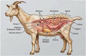
A visual, ex vivo examination was performed on the ovaries, oviducts, uterus, vagina, and urogenital sinus to identify pathologies and congenital anomalies. Measurements of the ovaries, functional structures, and other components were taken using a digital calliper (length and width) (Figure 3). The presence of paraovarian cysts, corpora lutea, and follicles was recorded through visual inspection. Measurements were used to confirm the presence of ovarian cysts, which were defined as follicles with a diameter greater than 10 mm. In the absence of follicles and/or corpora lutea, an ovary of small size (2 mm) was classified as inactive (Ngona et al., 2012). A paraovarian cyst was described as a cystic structure located on the ovary containing a clear fluid.
The incision of the general apparatus was performed to reveal the contents, which showed pathological uterine secretions of sanguinopurulent, mucopurulent, and purulent nature (Figure 4), and, where applicable, other pathological conditions in the remaining portions.
Statistical Analysis
Data were collected through surveys, observations, interviews, and sample collection. These were complemented by discussions with slaughterhouse operators and measurements of various structures from the sampled genital apparatus. The minimum sample size for six months was 97.4, equivalent to 97 specimens. We exceeded this size by 22%, obtaining a total sample size of 118. This approach provided a robust database processed and analysed using Microsoft Word and Microsoft Excel 2010, applying descriptive statistics.
RESULTS
Animals observed during data collection
The two main supply routes, Kasumbalesa and Kasenga, delivered animals whose age, based on dentition, ranged between 4AT and 8AT. However, individuals with 6AT were most frequently supplied (48.3 %) compared to those with 4AT (28.0 %) and 8AT (20.3 %). Health status assessed using the FAMACHA system showed individuals with normal ocular mucosa (pink) (58.5%), pale (23.7%), and dark (15.2%) (Table 2). Herd management in these supply routes indicated goats with weights mainly between 20 and 30 kg and ages corresponding to dentition stages from 4AT to 8AT. The Kasumbalesa and Kasenga routes provided most of the animals slaughtered, with weights generally in the good range (20–35 kg). The FAMACHA approach yielded results like those observed through weight assessment (pink conjunctiva). We classified goat weights into four categories: I: weight between 15 and 20 kg; II: weight between 20.1 and 25 kg; III: weight between 25.1 and 30 kg;
IV: weight between 30.1 and 35 kg.
Observation of ovarian activity through functional structures
Ovarian activity was assessed during data collection through ex vivo measurements of the ovaries. The respective mean values of length and width were 17.1 ± 2.72 mm and 11.7 ± 2.65 mm for the right ovary, and 17.5 ± 3.07 mm and 11.2 ± 2.69 mm for the left ovary. Functional structures provided a clearer illustration of this activity. Indeed, measurements of these structures confirmed the presence of corpora lutea and follicles. The length and width of the corpus luteum were, respectively, 11.1 ± 2.28 mm and 8.41 ± 1.86 mm (right ovary) and 11.3 ± 2.50 mm and 8.27 ± 2.09 mm (left ovary). Cavitary corpora lutea had smaller dimensions than non-cavitary ones, with respective length and width of 6.89 ± 0.91 mm and 5.70 ± 0.32 mm (right ovary) and 5.80 mm and 4.56 mm (left ovary). Follicles showed smaller values than corpora lutea, with length and width of 4.67 ± 1.30 mm and 3.42 ± 0.79 mm (right ovary) and 5.16 ± 1.59 mm and 3.91 ± 1.64 mm (left ovary).
Farms from the Kasumbalesa and Kasenga routes were more frequently involved compared to others. In these two routes, folliculogenesis was present in the glandular portion. Individuals aged between 4AT and 8AT, based on dentition, exhibited ovarian activity, particularly those with 6AT. Results showed an absence of folliculogenesis in individuals with 2AT. Females with pinkish conjunctiva displayed good ovarian activity.
Folliculogenesis was more pronounced in goats whose weights placed them in classes I, II, and III. Females in weight class IV exhibited low follicular activity. Specifically, females in classes I to III had a higher number of follicles (11–14 follicles) compared to those in class IV (2 follicles). However, the luteal phase was observed in females across all weight classes.
Classes II and III included females with strong ovarian activity during the luteal phase, with 51 and 96 corpora lutea counted, respectively. Classes I and IV showed lower numbers (22 and 21 corpora lutea, respectively).
Among females observed in Kamalondo slaughterhouses, 41.5% had corpora lutea at an age corresponding to 6AT (49 corpora lutea). Overall, corpora lutea were frequently observed in females aged between 4AT and 8AT. Females with 2AT dentition showed a low frequency of corpora lutea (4 corpora lutea). Females with pink conjunctiva had the highest number of corpora lutea (52.5%), while those with pale and dark conjunctiva had 22.9% and 11.9%, respectively.
Identification of pathologies in the glandular portion
A low frequency of paraovarian cysts, follicular cysts, and ovarian tumours was observed in females from Kamalondo slaughterhouses. The numbers recorded were 3, 3, and 4, respectively. According to origin, goats from Kasumbalesa and Kasenga exhibited follicular and paraovarian cysts. Ovarian tumours were observed in females from Kasenga and Kipushi (Table 3).
Females from the Kasenga and Kasumbalesa routes exhibited paraovarian cysts. Females from other routes showed no paraovarian cysts. Likewise, females whose age corresponded to dentition stages 2AT and 8AT did not present any. The same applied to females with pink or light ocular mucosa. Paraovarian cysts were present on both the left and right ovaries. Follicular cysts were rarely observed in goats slaughtered at Kamalondo (n = 3; 2.54%) and were associated with dentition stage 6AT. In terms of laterality, they were observed on the right ovary. Ovarian tumours were also infrequent (1.69%) and occurred in females with 6AT dentition.
Pathologies of the Tubular Portion and Urogenital Sinus
Four goats from the Kasumbalesa and Kasenga routes, aged according to dentition stages 4AT and 6AT, exhibited pathological uterine contents. Furthermore, based on the FAMACHA system, females with light and pink ocular mucosa were affected by these pathological contents, which were mucopurulent and sanguinopurulent in nature. Among the 118 samples, no subject presented pathologies in the urogenital sinus.
DISCUSSION
Animals Observed During Data Collection
Herd management in the different supply routes revealed individuals with weights generally ranging between 20 and 30 kg and ages corresponding to dentition stages from 4AT to 8AT. The Kasumbalesa and Kasenga routes provided most of the animals slaughtered, with weights generally in the good range. The correlation between age and weight varies according to goat breeds (Alexandre et al., 2012). This was observed in Djallonké goats in Chad. This meat-type breed showed a different age-weight correlation compared to goats in Lubumbashi. Djallonké goats exhibited increasing weight with age (13.5 kg at 1–2 years; 14.7 kg at 2–3 years; 18.2 kg at 3–4 years; 20 kg above 4 years) (Djagba et al., 2019). In contrast, goats observed in Lubumbashi displayed weight classes that did not allow precise age determination based on weight.
Health status assessed using the FAMACHA system showed individuals with varying ocular mucosa scores. However, females with a normal score (pink) were predominant (58.5 %), while extreme cases were also observed (pale conjunctiva: 23.7 %; dark conjunctiva: 15.2 %). Messager (2018) reported similar results for pink conjunctiva (49.8%) but different for dark (46.5%) and pale (3.3%). The FAMACHA system, widely used in sub-Saharan and southern Africa, provides a reliable assessment of female health status (Miller and Waller, 2004). This approach is highly useful since reproductive management depends on females with a positive energy balance. A negative energy balance leads to ovarian inactivity, resulting in subfertility and, subsequently, acquired sterility (Gnanda et al., 2008).
Observation of Ovarian Activity through Functional Structures
Ovarian activity was assessed ex vivo through ovary measurements. The values obtained corroborated those reported for goats in Lubumbashi (Ngona et al., 2012), with lengths and widths of 18 ± 0.43 mm and 13 ± 0.27 mm (left ovary) and 17 ± 0.41 mm and 13 ± 0.27 mm (right ovary). The results observed in Kamalondo slaughterhouses confirmed the absence of ovarian inactivity. This positive finding highlights the importance of reproductive management to ensure normal fertility (Pannetier et al., 2003). Follicle length and width were 4.67 ± 1.3 mm and 3.42 ± 0.79 mm (right ovary) and 5.16 ± 1.59 mm and 3.91 ± 1.64 mm (left ovary). These values, measured using a digital calliper, were lower than those reported by Moussa et al. (2019) for Sahel goats, where mean diameters measured by ultrasonography were 7.4 ± 2.23 mm (right ovary) and 7.13 ± 2.42 mm (left ovary). In Egyptian goats, follicle diameters averaged 8.0 ± 0.1 mm (right ovary) and 7.8 ± 0.2 mm (left ovary) (Simões et al., 2006). Follicles and corpora lutea are critical since ovulation results from follicular growth and maturation, which determine fertility potential (Zongo, 2015). Ovarian activity leads to the luteal phase, essential for gestation and, consequently, normal fecundity. Monitoring ovarian activity ensures herd reproductive management that promotes profitability (Moussa et al., 2019).
Identification of Pathologies in the Glandular Portion
Results from females observed in Kamalondo slaughterhouses showed a lower frequency of paraovarian cysts (5.22%) and higher frequencies for follicular cysts (0.65%) and ovarian tumours (0.65%) compared to a study on 153 dwarf goats in Algeria (Benchaib, 2007). Gustafsson and Holmberg (1996) reported a frequency of 0.6% for ovarian cysts in 502 genital organs examined ex vivo. This disorder, linked to ovarian cysts, often occurs before the first ovulation following postpartum. Many females will resume normal ovarian cycles, but the condition may persist if untreated (Hanzen, 2005). This hormonal disorder leads to follicular cyst formation, originating from an unruptured ovarian follicle whose ovum closes before undergoing cystic transformation (Felberg et al., 1989).
Pathologies of the Tubular Portion and Urogenital Sinus
Pathological uterine contents observed in Lubumbashi slaughterhouses were mucopurulent (0.84%) and sanguinopurulent (2.54%). These conditions were associated with puerperal metritis or clinical endometritis. Such pathologies generally follow cervico-vaginal inflammation but may also result from abortion or parturition (Arthur et al., 1992; Erer, 1995). The urogenital sinus showed no pathological changes in Lubumbashi slaughterhouses, consistent with findings by Benchaib (2007) in Algerian female goats.
CONCLUSION
The goats slaughtered in the Kamalondo municipality slaughterhouses mainly originated from the Kasumbalesa, Kasenga, Kipushi, Kinsever, Pweto, Bukama routes and neighborhoods within Lubumbashi city. The Kasumbalesa and Kasenga routes supplied more goats than the other routes. The weights of these goats varied (15 to 35 kg) and corresponded to ages with dentition stages of 4AT (28.0%) and 6AT (48.3%). According to the FAMACHA approach, 58.5% of these goats exhibited pink ocular mucosa. Measurements of the ovaries and the presence of functional structures demonstrated ovarian activity in goats slaughtered in Kamalondo. Some pathologies were observed in the glandular portion (follicular cysts, paraovarian cysts, ovarian tumours) and the tubular portion (mucopurulent and sanguinopurulent contents).
In perspective, reproductive management in goats is likely to undergo significant improvement to positively enhance numerical productivity in traditional goat farming, thereby increasing profitability. A preliminary study based on the origin of goats is recommended to ensure proper management of breeding females by farmers.
REFERENCES
Alexandre G., Arquet R., Fleury J., Troupé W., Boval M., Archimède H., Mahieu M., Mandonnet M. (2012). Systèmes d’élevage caprins en zone tropicale: analyse des fonctions et des performances. Inra prod. Anim., 25: 305-316.
Arthur G.H., Noakes D.E., Pearson H. (1992). Veterinary reproduction and obstetrics. 6th ed., 641 pages, bailliere tindall, london.
Benchaib F. (2007). Étude comparative, descriptive et diagnostique de la pathologie genitale chez la femelle des petits ruminants en algérie. Universite d’oran -Es-Senia, faculté des sciences département de biologie, 163pp. 52-87.
Bousquet C.A. (2005). Pathologie caprine en deux-sèvres: état des lieux et impact sur les niveaux de réforme et de mortalité. Thèse d’agrégation, 154p, 90-111.
Casamitjana P. (1998). Facteurs d’infertilité chez les petits ruminants. In journées nationales des GTV: La reproduction, tours, 27-29 mai 103p. 44, 57-60
Djagba A.Y., Bonfoh B., Dayo G.K., Aklikokou K. A., Bassowa H. (2019). Variabilité des caractères morphologiques mesurables de la chèvre Djallonké dans les zones agro-écologiques du Togo. Tropicultura, 37(2).
Erer S. (1995). Clinical aspects of endometritis and metritis in small ruminants. Veterinary Journal, 150: 245252.
Felberg D., Yeshaya A., Ashkenazi J., Goldman G.A., Dicker D., Goldman J.A. (1989). Ovarian cyst formation: A complication of gonadotrophin releasing hormone agonist therapy. Fertil. Steril., 51: 4245.
Frette F., Humbert G. (2022). Fédération nationale des éleveurs de chèvres, la préparation des chèvres à la réforme. 5p: 2.
Gnanda B.I., Zoundi S.J., Nianogo J.A., Meyer C., Zono O. (2005). Test d’un complément minéral et azoté sur les paramètres de reproduction de la chèvre du sahel burkinabé. Revue élev. Méd. Vét. Pays trop., 4: 257-265.
Gustafsson B., Holmberg O. (1996). Postmortem examination of the genital tract in ewes with special reference to the occurrence of developmental abnormalities. Svensk. Vet. - Tidn., 18: 432-436.
Hadjé M. (2014). État des lieux des abattoirs et aires d’abattage situés dans trois regions du Tchad. Doctoral dissertation, University Cheikh Anta Diop de Dakar.
Hanzen C. (2005). L’infertilité bovine, approche individuelle ou de troupeau ? Le point vétérinaire, 84: 84-88.
INRA France (1999). Implications économiques des pratiques de réforme et de renouvellement dans les grands troupeaux caprins laitiers. Renc. Rech. Ruminants, 6: 39-42.
Jansen C., Kees V.D.B. (2004). Agrodok 7: l’élevage de chèvres dans les zones tropicales, 103 p.
Kalenga H.K., Vandenput S., Antoine N., Moula N., Kashala J-C.K., Farnir F., Le Roy P. (2015). Amélioration génétique caprine à Lubumbashi (RDC): Analyse ACP de mensurations de la population locale. Livestock research for rural development, 27: 234.
Le Guillou S. (1998). Reproduction et alimentation chez les petits ruminants. In journées nationales des gtv: La reproduction. 98p.
Messager A.B. (2018). Etude épidémiologique du parasitisme influençant la production de cachemire à Bayankhongor (mongolie) et propositions de solutions adaptées dans le cadre d’une production durable. Doctoral Dissertation, École nationale vétérinaire d’Alfort 128p.
Miller J.,Waller P. (2004). Novel approaches to control of parasites – a workshop. Vet. Parasitol., 125: 59-68.
Moussa A., Traoré M., Diallo S., Konaté B. (2019). Follicular dynamics and ovarian activity in Sahel goats under tropical conditions. Animal Reproduction Science, 207: 4552.
Mujinga W., Lwamba J., Mutala S., Hüsken S.M.C. (2009). Inventaire des espèces de poisson disponibles sur les marchés urbains de Lubumbashi, République Démocratique du Congo. Novembre.
Ngona I.A. (2008). Performances et facteurs d’influence de la reproduction de l’espèce caprine en milieu tropical. Thèse d’agrégation, faculté de médecine vétérinaire, université de Lubumbashi. 165 p.
Ngona I.A., Beduin J.M., Khang’maté A.B.F., Hanzen C. (2012). Etude descriptive des caractéristiques morphométriques et génitales de la chèvre de Lubumbashi en République Démocratique du Congo. Revue d’élevage et de médecine vétérinaire des pays tropicaux, 65 : 75-79.
Ngona I.A., Masenga P., Khang’maté A.B. (2015). La gestion de la reproduction caprine au travers des femelles vendues à différents points de vente de Lubumbashi en République Démocratique du Congo. Journal of Applied Biosciences, 91: 8511-8518.
Pannetier M., Servel N., Cocquet J., Besnard N., Cotinot C., Pailhoux E. (2003). Expression studies of the pis regulated genes suggest different mechanisms of sex determination within mammals. Cytogenetic and genome research, 101: 199-205.
Poncelet J.L. (2004). Dentisterie: chronologie de l’évolution de la dentition (diagnose de l’âge) 3p.
Simões J., Almeida J.C., Valentim R., Baril G., Azevedo J., Fontes P., Mascarenhas R. (2006). Follicular dynamics in serrana goats. Anim. Reprod. Sci., 95: 16-26.
Useni S.Y., Malaisse F., Kaleba S.C., Kankumbi F.M., Bogaert J., (2017). Le rayon de déforestation autour de la ville de Lubumbashi (HautKatanga, RD Congo): Synthèse. Tropicultura, 35: 67.
Weather S., (2021). Les climats de la RD Congo: https://fr.weatherspark.com/y/94251/météo-moyenne-à-lubumbashi-congo-kinshasa-tout-au-long-de-l’année.
Whitney K.M., Valentine B.A., Schlafer D.H. (2000). Caprine genital leiomyosarcoma. Veterinary Pathology, 37: 89-94.
Zarechny M.V. (2020). Détermination de l’âge chez les chèvres par la dentition https://garden-fr.desigusxpro.com/kozy/kak-opredelit-vozrast-metody.html.
Zongo M. (2015). Application de l’échographie à la reproduction de la chèvre du sahel. Thèse doctorat, Université Ouaga I, Ouagadougou.